Pseudopelade of Brocq, often referred to simply as “pseudopelade,” is an uncommon and somewhat enigmatic form of scarring (cicatricial) alopecia primarily affecting the scalp. First described by Louis-Anne-Jean Brocq in the late 19th century, it is characterized by patchy areas of permanent hair loss that develop gradually and usually without overt signs of inflammation. Unlike other forms of cicatricial alopecia, pseudopelade of Brocq often presents with very subtle changes in the scalp, making it challenging to diagnose in its early stages. This article aims to provide a comprehensive overview of pseudopelade of Brocq at an MSc level, discussing epidemiology, clinical presentation, pathophysiology, diagnosis, and treatment approaches.
Historical Context and Nomenclature: Pseudopelade of Brocq derives its name from the original description by Brocq, who observed patchy alopecia resembling “pelade” (the French term for alopecia areata), but failing to fit precisely within the well-known patterns of alopecia areata or other clearly defined forms of hair loss.
Over the years, controversy has surrounded this nomenclature, as dermatologists and hair researchers have debated whether pseudopelade of Brocq truly represents a distinct entity or rather a late-stage manifestation of other scarring alopecias such as lichen planopilaris, discoid lupus erythematosus, and/or folliculitis decalvans. While debates persist, many in the field consider pseudopelade to be an end-stage, “burned-out” presentation of various inflammatory processes culminating in the same clinical and histopathological outcome: scarring and permanent hair loss.
Nevertheless, the term “pseudopelade of Brocq” remains widely used in clinical practice to describe scarring alopecia that presents with minimal visible inflammation and leaves behind smooth, irregular patches of hair loss. A better understanding of its etiology and pathogenesis continues to evolve with advancements in dermatological research and more refined histopathological techniques.
Epidemiology: Pseudopelade of Brocq is relatively rare compared to other scarring alopecias, with precise incidence and prevalence data difficult to establish due to overlaps with conditions of similar clinical presentation. Epidemiological data indicate no clear gender predilection in some studies, while others suggest it may be slightly more common in middle-aged women. Because of the subtleness of the disease, many cases likely go undiagnosed or are diagnosed late, further complicating epidemiological assessments.
Age of onset can range broadly, from young adulthood to late middle age, though most patients notice the first signs of patchy alopecia between 30 and 50 years of age. There does not appear to be a strong familial or genetic component clearly linked to pseudopelade of Brocq, differentiating it from some other scarring alopecias that can sometimes cluster within families.
Clinical Presentation: The hallmark feature of pseudopelade of Brocq is the gradual development of small, discrete, asymptomatic patches of hair loss on the scalp. These patches often have the following characteristics:
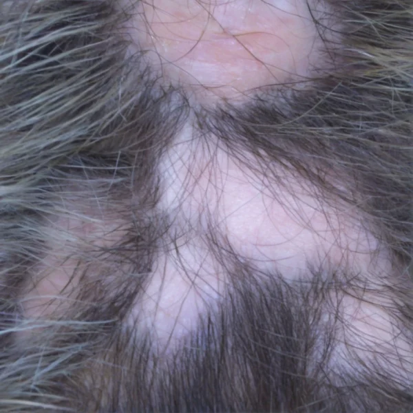
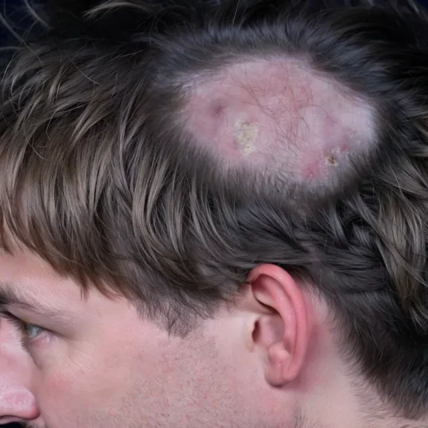
Smooth, Atrophic Patches: Affected areas of the scalp typically appear pale, smooth, and slightly depressed or atrophic. There is usually an absence of the visible scale or erythema commonly seen in more classic inflammatory scarring alopecias like lichenplanopilaris.
Minimal Inflammation: Patients often report little to no itching, burning, or discomfort. On close clinical inspection, overt signs of inflammation – such as redness or follicular hyperkeratosis – are either subtle or absent.
Varied Patch Shapes: Lesions can be round, oval, or irregular in outline. The shape can be quite distinct from the smooth, clearly demarcated patches of alopecia areata.
Slow Progression: Hair loss typically occurs over a long duration. Patients may not notice any sudden changes, given the generally asymptomatic nature of the process.
The distribution on the scalp can vary widely, though many patients present with patches on the vertex or parietal regions. Over time, the patches may coalesce, leading to large areas of confluent alopecia. Occasionally, isolated “islands” of hair persist within these patches, accentuating the irregular, “footprint in the snow” appearance commonly described in pseudopelade.
Pathophysiology: Despite more than a century of clinical observations, the precise pathophysiological mechanism underlying pseudopelade of Brocq remains incompletely understood. Histologically, it manifests as a primary lymphocytic cicatricial alopecia, characterized by:
Lymphocytic Infiltrate: Microscopic examination of scalp biopsies often shows a predominantly lymphocytic infiltrate around the hair follicles, similar in some respects to what is seen in lichen planopilaris or discoid lupus erythematosus, yet usually less intense.
Follicular Destruction: Progressive destruction of the follicular units results in permanent hair loss. Over time, the inflammation “burns out,” leaving little inflammatory infiltrate in later stages. This end-stage scarring leads to complete replacement of the hair follicle by fibrotic tissue.
Epithelial Atrophy: Hair follicles in the affected areas show miniaturization and atrophy of the follicular epithelium, contributing to the characteristic atrophic, smooth scalp appearance.
One theory is that pseudopelade of Brocq could represent the terminal stage of multiple inflammatory pathways that converge in follicular scarring. The inflammation around the isthmus and infundibulum of hair follicles compromises the follicular stem cells, ultimately leading to irreversible alopecia.
Differential Diagnosis: Diagnosing pseudopelade of Brocq requires careful consideration of other scarring alopecias. Important differential diagnoses include:
Lichen Planopilaris (LPP): LPP is a common cause of primary lymphocytic cicatricial alopecia, usually characterized by perifollicular erythema, scale, and pruritus. Over time, it can appear clinically similar to pseudopelade if inflammation subsides.
Discoid Lupus Erythematosus (DLE): DLE often presents with well-demarcated plaques, scaling, and dyspigmentation, but late-stage scarring can mimic pseudopelade. Histopathological evaluation can help identify lupus-specific features.
Alopecia Areata (Scarring Variant): Classical alopecia areata is typically non-scarring, but a rare scarring variant may occasionally resemble pseudopelade, although inflammation and histopathology usually differ.
Central Centrifugal Cicatricial Alopecia (CCCA): Commonly seen in women of African descent, CCCA begins at the vertex and expands outwards. Early inflammatory signs (itch, burning) are more prominent than in pseudopelade, but late-stage scarring may overlap in appearance.
Folliculitis Decalvans: This neutrophilic form of cicatricial alopecia usually presents with pustules and crusts, which are not typical in pseudopelade. Nonetheless, end-stage scarring can sometimes appear similar.
Given these overlaps, a definitive diagnosis of pseudopelade of Brocq often hinges on scalp biopsy findings combined with clinical correlation.
Diagnosis: The diagnostic process for pseudopelade of Brocq involves a thorough medical history, physical examination, trichoscopic evaluation, and histopathological analysis. Key steps include:
Clinical Assessment: Dermatologists assess the scalp for lesion distribution, morphology, presence of erythema, scaling, and any sign of inflammation. The absence or minimal presence of symptomatic inflammation is a clinical clue pointing towards pseudopelade.
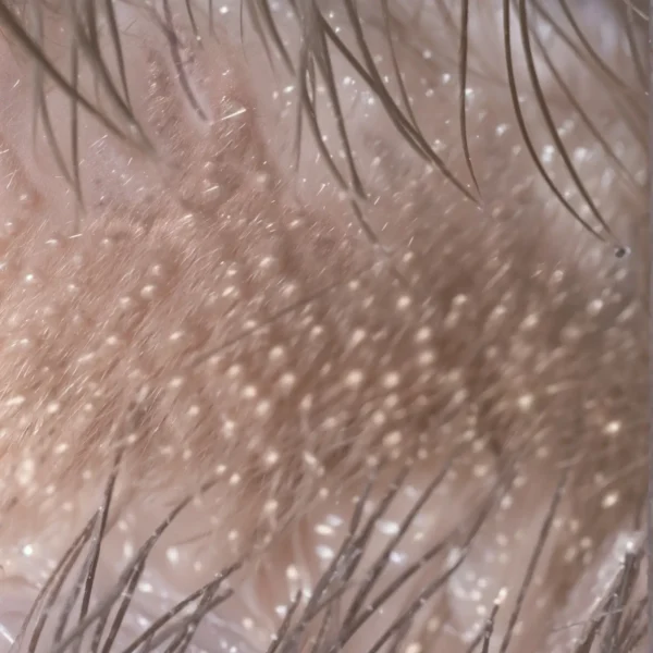
Trichoscopy: Using a dermatoscope, clinicians may observe features such as subtle perifollicular erythema, reduced follicular ostia, and the presence of lonely hair strands in atrophic areas (“lonely hair sign”). While these findings are not pathognomonic, they provide supportive evidence for cicatricial alopecia.
Scalp Biopsy: A punch biopsy from an active or borderline lesion is often vital to diagnosis. Histopathological analysis can reveal a primary lymphocytic infiltrate targeting the upper portion of the follicle, along with fibrous tract formation replacing the destroyed follicles. In early stages, some inflammatory changes may still be present, whereas end-stage lesions often display little to no active inflammation, reflecting a “burned-out” process.
Laboratory Tests: Basic screening tests for autoimmune markers (e.g., antinuclear antibodies) can help exclude lupus erythematosus. Additional tests can be carried out depending on clinical suspicion.
Arriving at a definitive diagnosis can be challenging, and sometimes pseudopelade of Brocq remains a diagnosis of exclusion, especially when patients present in advanced stages with nonspecific scarring.
Management and Treatment Approaches: Given the subtle progression of pseudopelade of Brocq and the lack of overt inflammation, treatment options generally focus on halting disease progression, alleviating any subclinical inflammation, and preserving residual hair follicles as far as possible. Key management strategies include:
Topical Corticosteroids: High-potency topical corticosteroids or intralesional steroid injections may help suppress underlying lymphocytic inflammation, particularly in early or moderately active disease. Although overt inflammation is minimal, the pathophysiology still involves immune-mediated damage.
Topical Calcineurin Inhibitors: Agents such as tacrolimus or pimecrolimus can be used off-label to reduce lymphocytic infiltration around hair follicles. Their effectiveness varies, and they tend to be more beneficial in inflammatory stages.
Systemic Therapies: In cases where multiple patches are progressing, systemic immunomodulatory drugs such as oral corticosteroids, hydroxychloroquine, or methotrexate may be considered to help dampen the immune response. The choice of therapy often depends on the patient’s disease activity, comorbidities, and tolerance of side effects.
Minoxidil: While minoxidil cannot reverse scarring, it may help strengthen existing, non-scarred hair follicles around affected areas, potentially slowing progression or improving the cosmetic appearance.
Laser and Light-Based Therapies: Limited evidence exists on the role of low-level laser therapy (LLLT) or photodynamic therapy in pseudopelade of Brocq. Some anecdotal reports suggest mild improvements in hair density, but the scarring nature of the condition ultimately limits the efficacy of these modalities.
Surgical Options: Hair transplantation is generally not considered for active scarring alopecias. However, in stable, long-standing disease where the inflammatory process has clearly burned out, hair restoration surgery may be an option to improve cosmetic appearance, provided there is sufficient donor hair and no ongoing signs of active follicular destruction.
Supportive Measures: Psychological and social support are important for patients coping with the visible aspects of hair loss. Camouflaging techniques (e.g., scalp micropigmentation, hairpieces) and counseling can significantly improve quality of life.
Ultimately, the primary goal is to prevent disease progression rather than to recover lost hair, as scarring alopecias are fundamentally irreversible once follicles are destroyed.
Prognosis: The clinical course of pseudopelade of Brocq can be unpredictable. While some patients experience a very slow progression that eventually “burns out” with only limited patches of hair loss, others may see a more extensive pattern of scarring alopecia over time. Once the condition stabilizes and no active inflammation remains, further hair loss typically ceases, although regrowth in scarred areas is not possible.
Early detection and proactive management of subclinical inflammation can potentially limit the extent of permanent hair loss. Patients must be followed periodically to assess disease progression, even if the scalp appears quiet, as subtle inflammation can persist beneath the surface.
Future Directions and Research: Ongoing research continues to probe the immunological and molecular underpinnings of scarring alopecias, including pseudopelade of Brocq. Advances in genetic profiling, stem cell biology, and immunomodulatory treatments may eventually offer improved diagnostic markers and more targeted therapeutic strategies. Specifically:
Biomarker Identification: Further studies are necessary to isolate biomarkers that could definitively distinguish pseudopelade of Brocq from lichen planopilaris or lupus-associated cicatricial alopecias, streamlining diagnostic accuracy.
Regenerative Medicine: Experimental therapies involving growth factors, platelet-rich plasma (PRP), or stem cell technologies hold promise. Although scarring alopecia is difficult to reverse, localized stem cell therapies might one day stimulate partial regrowth or prevent progression.
Immunotherapy: Building on the success of biologic agents in other inflammatory skin diseases, future investigations may develop monoclonal antibodies or small-molecule inhibitors that selectively modulate the pathological lymphocytic attack on hair follicles in pseudopelade.
A better understanding of the condition’s root causes will allow clinicians to tailor treatments more precisely and perhaps alter the disease course rather than simply slow it.
Conclusion: Pseudopelade of Brocq remains a challenging and somewhat vague form of primary scarring alopecia. It presents with subtly progressive, patchy hair loss, often escaping early detection due to minimal inflammation and a relatively asymptomatic course. Although controversies persist about whether pseudopelade is a distinct entity or an end-stage variant of other scarring alopecias, it is clinically recognized for its characteristic smooth, footprint-like patches of permanent alopecia.
Diagnosis relies heavily on careful clinical examination, trichoscopic findings, and histopathological evaluation from scalp biopsies. Treatment aims to arrest disease progression through immunomodulatory therapies, although no single regimen is universally effective. Patients often benefit from a multidisciplinary approach, integrating medical management, psychosocial support, and, in select cases, surgical intervention once the disease is inactive.
As our understanding of the inflammatory mechanisms and stem cell biology underlying pseudopelade of Brocq evolves, we can hope for more targeted and effective therapies in the future. For patients dealing with the psychological impact of scarring alopecia, a comprehensive treatment plan that addresses both the physical and emotional aspects of hair loss remains indispensable. By combining early diagnosis, individualized therapy, and continued research into its pathogenesis, the dermatological community aims to improve outcomes and quality of life for those affected by this complex condition.
Gay Prieto J. Pseudopelade of Brocq: its relationship to some forms of cicatricial alopecias and to lichen planus. J Invest Dermatol. 1955 Mar;24(3):323–35.
1.
Braun-Falco O, Imai S, Schmoeckel C, Steger O, Bergner T. Pseudopelade of Brocq. Dermatologica. 1986;172(1):18–23.
1.
Pierard-Franchimont C, Pierard GE. Massive lymphocyte-mediated apoptosis during the early stage of pseudopelade. Dermatologica. 1986;172(5):254–7.
1.
Pincelli C, Girolomoni G, Benassi L. Pseudopelade of Brocq: an immunologically mediated disease? Dermatologica. 1987;174(1):49–50.
1.
Dawber R. What is pseudopelade? Clin Exp Dermatol. 1992 Sep;17(5):305–6.
1.
Silvers DN, Katz BE, Young AW. Pseudopelade of Brocq is lichen planopilaris: report of four cases that support this nosology. Cutis. 1993 Feb;51(2):99–105.
1.
Madani S, Trotter MJ, Shapiro J. Pseudopelade of Brocq in beard area. J Am Acad Dermatol. 2000 May;42(5 Pt 2):895–6.
1.
Amato L, Mei S, Massi D, Gallerani I, Fabbri P. Cicatricial alopecia; a dermatopathologic and immunopathologic study of 33 patients (pseudopelade of Brocq is not a specific clinico-pathologic entity). Int J Dermatol. 2002 Jan;41(1):8–15.
1.
Sakamoto F, Ito M, Saito R. Ultrastructural study of acquired pili torti-like hair defects accompanying pseudopelade. J Dermatol. 2002 Apr;29(4):197–201.
1.
Moretti S, Amato L, Massi D, Bianchi B, Gallerani I, Fabbri P. Evaluation of inflammatory infiltrate and fibrogenic cytokines in pseudopelade of Brocq suggests the involvement of T-helper 2 and 3 cytokines. Br J Dermatol. 2004 Jul;151(1):84–90.
1.
Alzolibani AA, Kang H, Otberg N, Shapiro J. Pseudopelade of Brocq. Dermatol Ther. 2008;21(4):257–63.
1.
Otberg N, Wu WY, McElwee KJ, Shapiro J. Diagnosis and management of primary cicatricial alopecia: part I. Skinmed. 2008;7(1):19–26.
1.
Yu M, Bell RH, Ross EK, Lo BKK, Isaac-Renton M, Martinka M, et al. Lichen planopilaris and pseudopelade of Brocq involve distinct disease associated gene expression patterns by microarray. J Dermatol Sci. 2010 Jan;57(1):27–36.
1.
Kittridge A, Haught JM, English JC. Alopecia areata mimicking pseudopelade of Brocq. Cutis. 2010 Oct;86(4):187–9.
1.
Harries MJ, Paus R. The pathogenesis of primary cicatricial alopecias. Am J Pathol. 2010 Nov;177(5):2152–62.
1.
Ghosh S, Jain VK. “Pseudo” Nomenclature in Dermatology: What’s in a Name? Indian J Dermatol. 2013 Sep;58(5):369–76.
1.
Baeza-Hernández G, Jaquero-Valero MI, Rubio-Aguilera RF, Araya-Umaña LC, Horcajada-Reales C, Moreno-Torres A. Elastic Stain in Pseudopelade of Brocq: a Helpful Histopathological Diagnostic Clue. Dermatol Pract Concept. 2023 Apr 1;13(2):e2023088.
Introduction: Keratosis Follicularis Spinulosa Decalvans (KFSD), also referred to as Keratosis pilaris decalvans, is a rare genodermatosis characterized by progressive follicular keratosis, leading to scarring…
Scarring alopecias, sometimes also known as cicatricial alopecias, represent a heterogeneous group of disorders that result in irreversible hair loss due to the destruction of…
Manage Cookie Consent
We use technologies like cookies to store and/or access device information. We do this to improve browsing experience and to show (non-) personalized ads. Consenting to these technologies will allow us to process data such as browsing behavior or unique IDs on this site. Not consenting or withdrawing consent, may adversely affect certain features and functions.
Functional Always active
The technical storage or access is strictly necessary for the legitimate purpose of enabling the use of a specific service explicitly requested by the subscriber or user, or for the sole purpose of carrying out the transmission of a communication over an electronic communications network.
Preferences
The technical storage or access is necessary for the legitimate purpose of storing preferences that are not requested by the subscriber or user.
Statistics
The technical storage or access that is used exclusively for statistical purposes.The technical storage or access that is used exclusively for anonymous statistical purposes. Without a subpoena, voluntary compliance on the part of your Internet Service Provider, or additional records from a third party, information stored or retrieved for this purpose alone cannot usually be used to identify you.
Marketing
The technical storage or access is required to create user profiles to send advertising, or to track the user on a website or across several websites for similar marketing purposes.