Hair, an essential aspect of human aesthetics and identity, is far more complex than its simple appearance suggests. Each strand of hair is a marvel of biological engineering, with distinct layers and structures contributing to its overall health, strength, and appearance. For large terminal hair follicles, such as those on the scalp, the hair shaft is composed of three parts. From the outermost to innermost part of a hair fiber, it is comprised of the cuticle, the cortex, and the medulla in the center. This article delves into the intricate world of hair fiber structure, particularly focusing on the cuticle layer as it forms and develops within the hair follicle. Understanding the hair cuticle and its formation is crucial for comprehending the dynamics of hair care, damage, and repair.
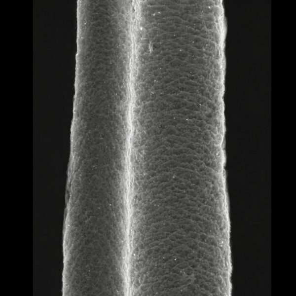
Hair Fiber Cuticle: The human hair cuticle, which is the outermost layer of the hair shaft, is composed of multiple layers of cells. Typically, the cuticle consists of 5 to 10 overlapping layers of cells. Finer / smaller hair tends to have fewer cuticle layers. These layers resemble shingles on a roof and are responsible for protecting the inner regions of the hair fiber, particularly the cortex. The cuticle cells are flat and scale-like, and the health and condition of the cuticle layers are crucial for the overall integrity and appearance of the hair. Damage to these layers, which can occur through chemical processes, heat styling, or mechanical stress, can lead to hair that appears dull, is more prone to breakage, and has increased porosity. Maintaining the integrity of the cuticle is thus a key focus in hair care, emphasizing the importance of gentle treatment and protective hair care practices.
Unlike the cells of the inner root sheath (only found around the hair fiber inside the skin), the cuticle cells of the cortex do not show the presence of trichohyalin. Trichohyalin is a structural protein that is produced and retained in the cells of the inner root sheath and also in the medulla of a hair. For hair research scientists, distinguishing between the hair cuticle and the inner root sheath of a hair follicle is often done by looking for presence / absence of trichohyalin. The cells of the cuticle can also be differentiated from cells destined to become the hair fiber cortex by their lack of melanin (pigment). Using these features helps to identify the hair cuticle layer as it forms around the hair cortex of a hair fiber as it is being built inside a hair follicle.
The cells of the cuticle can be recognized in the upper part of the bulb as they sweep upward from the hair matrix. Initially the hair cuticle is just a single layer of cells and about midway up the hair follicle bulb, these cells are cuboidal in shape. When stained with a basic dye, the microscopic appearance of a histological section of these cells is found to be etched with numerous granules. When the cells reach the upper region of the hair bulb, they become columnar, with the long axis oriented radially. This orientation is maintained for a short distance above the bulb, and then their outer edges begin to be tipped upward. Since these cells are at least twice as broad as they are high, they progressively become more overlapping (imbricated) when the outer sides shift at the tips. Imbrication is a pattern or design having regularly arranged, overlapping edges, as roof tiles or fish scales.
As the cuticle cells’ orientation shifts from a horizontal plane to a vertical one, the cells become more and more flattened. This orientation is completed below the midway mark of the hair follicle. In the upper half of the follicle, these cells undergo hyalinization, a form of degeneration. Their nuclei disappear, and the mature cuticle becomes glued to the cells of the hair cortex. A major function of cuticle on the hair shaft is to protect the inner, softer tissue called the cortex, or else the cortex would become frayed and fall apart pretty quickly after emerging above the skin surface.
The cells of the cuticle of the hair are interlocked with those of the inner root sheath; this firmly anchors the hair in the follicle. The inner root sheath must grow at pretty much the same rate as the hair, as it molds and guides the hair shaft in its passage outward to the skin surface. When you pluck a hair fiber, the strength you need to pull it out essentially has to overcome the interlock between the hair cuticle and the inner root sheath, ripping the two apart. They are tightly interlinked, so it needs quite a lot of pull force to separate them.
The scale pattern of the cuticle in human hairs is routinely imbricate. Cuticles are also classified as coronal when each cell completely surrounds the hair. This can happen quite often with fine vellus hair, though it is much less likely that a single cell can reach all the way around a terminal sized hair fiber. More usually, there are several cuticle cells the overlap with each other to encircle terminal hair fibers. The free edge of cuticle cells may be simple, dentate (with short ridges), or serrate (saw-edged). Cuticle cells may be elongate, acuminate (gradually tapering to a point), ovate (egg shaped), or flattened.
The cuticle cells have different staining properties than the surrounding structures, which is likely due to the chemical differences in the keratins in cuticle cells. A different article elsewhere on keratin.com summarizes the differences in hair keratins in the hair fiber. The cuticle is responsible for much of the mechanical strength of the hair fiber. A healthy cuticle is more than just a protective layer, as the cuticle also represents the structure that controls the water content of the fiber. Much of the shine that makes healthy hair so attractive is due to the cuticle.
Hair fiber cortex: Cortical cells constitute the bulk of a hair, and it is the cortex that gives a hair fiber its eventual shape, resilience, elasticity and curl. The cortex as the main body of the hair is composed of elongated and fusiform (spindle-shaped) cells. Within each cortical cell are bundles of hair proteins called fibrils, running parallel to the fiber axis, and between the fibrils is a softer material called the matrix.
The cortex may contain cortical fusi, pigment granules, and/or large oval-to-round-shaped structures called ovoid bodies. Cortical fusi are irregular-shaped airspaces of varying sizes. They are commonly found near the root of a mature human hair, although they may be present throughout the length of the hair fiber. In the living portion of the hair root the fusi are filled with fluid; as the hair grows and dries out, air replaces the fluid. Pigment granules are small, dark, and solid structures that are granular in appearance and considerably smaller than cortical fusi. They vary in color, size, and distribution in a single hair.
Hair fiber medulla: In some of the largest terminal hairs in humans, the cortex has a central, relatively hollow core, called the medulla. The medulla is easily identified as a pale-staining, sometimes discontinuous line of cuboidal cells. The medulla may be as large as one third of the diameter of the hair fiber, and may be continuous, discontinuous, or fragmental. In coarse hairs it is usually continuous or fragmental, whereas in finer hairs it appears discontinuous or, it is absent altogether.
Some scientists and dermatologists are of the opinion that even though some adult hairs appear non-medullated when observed under a light microscope, when viewed under polarized light, all terminal hairs, with the exception of the finest ones (less than 60 micrometers diameter), show a fragmental or discontinuous medulla. The medulla may be only one or two cells thick, but it is present nonetheless.
In some animals, the air within the medulla helps to increase the insulating properties of the hair, contributing to the regulation of body temperature. Humans always have an amorphous medulla, if it’s present, and so it contains relatively less air than in hair medullae of other animals. To a large extent, intra and intercellular air spaces in the medulla determine the sheen and color tones of the hair by affecting the reflection of light. This is partly why hair color looks a lot different in sunlight than it does in the shade.
Conclusion: The detailed exploration of hair fiber structure, particularly the cuticle, cortex, and medulla, reveals the intricate and sophisticated nature of human hair. From the protective and overlapping layers of the cuticle to the strength and elasticity provided by the cortex, and the unique properties of the medulla, each component plays a vital role in the hair’s overall function and appearance. This knowledge is not only fundamental for scientists and dermatologists but also enlightening for anyone interested in the science behind hair care and health. Understanding these complex structures at a microscopic level can lead to better hair care practices, innovative treatments for hair and scalp diseases, and a deeper appreciation of the human body’s remarkable capabilities.
Montagna W, Van Scott EJ. The anatomy of the hair follicle. In: Montagna W, Ellis RA, editors. The Biology of Hair Growth. New York: Academic Press; 1958. p. 39–64.
1.
Hashimoto K. Ultrastructure of cuticle of human beard hair. In: The First Human Hair Symposium. New York: Medcom Press; 1974. p. 286–330.
1.
Hashimoto K. The structure of human hair. Clin Dermatol. 1988;6(4):7–21.
1.
Jones LN. Hair structure anatomy and comparative anatomy. Clin Dermatol. 2001;19(2):95–103.
1.
Steinert PM, Parry DAD, Marekov LN. Trichohyalin mechanically strengthens the hair follicle: multiple cross-bridging roles in the inner root shealth. J Biol Chem. 2003 Oct 17;278(42):41409–19.
1.
Khumalo NP. African hair morphology: macrostructure to ultrastructure. Int J Dermatol. 2005 Oct;44 Suppl 1:10–2.
1.
Takahashi T, Hayashi R, Okamoto M, Inoue S. Morphology and properties of Asian and Caucasian hair. J Cosmet Sci. 2006;57(4):327–38.
1.
Nagase S. Hair Structures Affecting Hair Appearance. Cosmetics. 2019 Sep;6(3):43.
Grooved hair, or pili canaliculi, is a rare and unusual condition where hair fibers exhibit longitudinal grooves running along their length. This condition can affect…
Circle hairs, also referred to as spiral hairs, represent a fascinating and somewhat under-discussed topic within the domain of dermatology, specifically within the study of…
Manage Cookie Consent
We use technologies like cookies to store and/or access device information. We do this to improve browsing experience and to show (non-) personalized ads. Consenting to these technologies will allow us to process data such as browsing behavior or unique IDs on this site. Not consenting or withdrawing consent, may adversely affect certain features and functions.
Functional Always active
The technical storage or access is strictly necessary for the legitimate purpose of enabling the use of a specific service explicitly requested by the subscriber or user, or for the sole purpose of carrying out the transmission of a communication over an electronic communications network.
Preferences
The technical storage or access is necessary for the legitimate purpose of storing preferences that are not requested by the subscriber or user.
Statistics
The technical storage or access that is used exclusively for statistical purposes.The technical storage or access that is used exclusively for anonymous statistical purposes. Without a subpoena, voluntary compliance on the part of your Internet Service Provider, or additional records from a third party, information stored or retrieved for this purpose alone cannot usually be used to identify you.
Marketing
The technical storage or access is required to create user profiles to send advertising, or to track the user on a website or across several websites for similar marketing purposes.